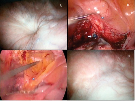
Fig. 1.

Laparoscopic and cystoscopic images. (A) The cystoscopic view of the 0.5-mm orifice of fistula at the bladder dome. (B) Methylene blue leakage from the bladder after excision of the fistula tract (white arrow). The asterisk shows bladder. (C) The mobilization of the peritoneum of the anterior abdominal wall with a transverse incision. The asterisk shows the mobilized peritoneal flap. (D) Postoperative control cystoscopy revealed well-healed scar tissue at the bladder dome.
