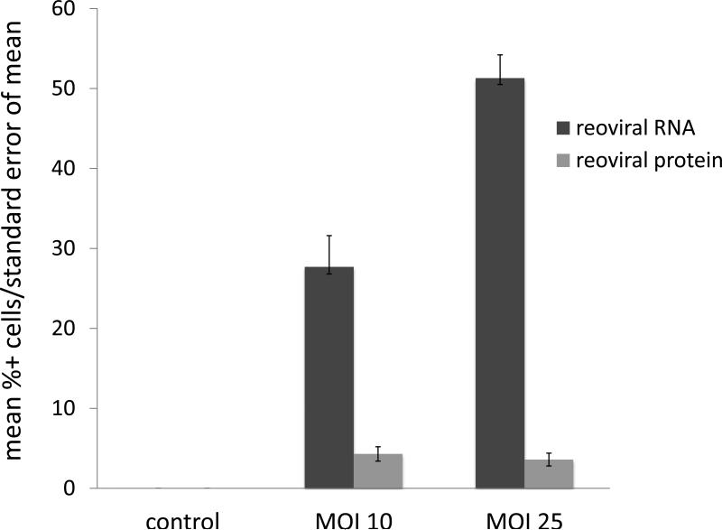
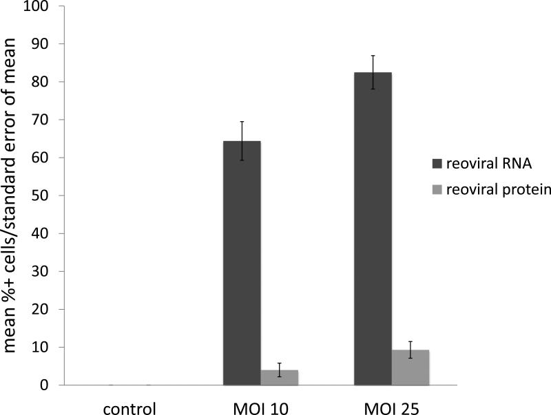
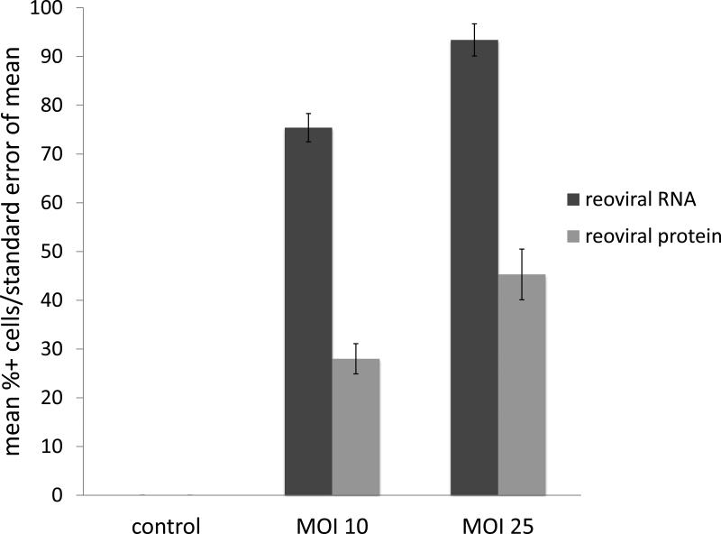
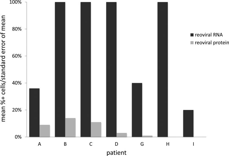
Figure 1. Reoviral RNA and capsid protein expression.
Reoviral RNA was present at multiplicity of infection (MOI) 10 and 25 in resistant (OPM2), intermediately resistant (H929), and sensitive (RPMI-8226) MM cell lines (1a – 1c). Viral RNA and protein increased with increased MOI. Reoviral protein was highest in the sensitive MM cell line RPMI-8226. Seven patients had adequate tissue present after Reolysin treatment for analysis of reoviral RNA and capsid protein expression (1d). Each value represents the percentage of CD138+ cells that were found to have co-localization of reoviral RNA or protein and CD138+. Results indicate that viral RNA was present in a much higher percentage of CD138+ cells than reoviral protein, suggesting that viral entry was present but replication was minimal.


