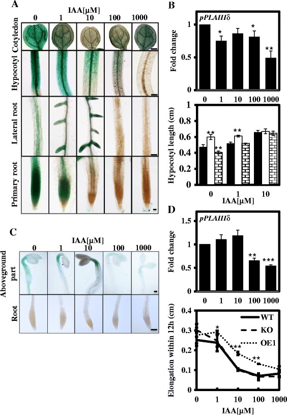
Figure 8.

Response of pPLAIIIδ to exogenous IAA induction. (A) GUS activity in 7-day-old transgenic Arabidopsis plants carrying pPLAIIIδ:GUS fusions under treatment with 0 to 1 mM IAA for 48 h. The intensity of GUS staining decreased gradually with increases in the exogenous IAA concentration in multiple organs, except for the sites of the lateral root initiation under 10 μM. Cotyledon, hypocotyl and lateral root, Bar = 500 μm; Primary root, Bar = 100 μm. (B) Hypocotyl length of WT, KO and OE1 plants incubated with 0 to 10 μM IAA in the light. The transcript levels of pPLAIIIδ in the light were quantified following incubation with 0 to 1000 μM IAA via real-time PCR, using ACT7 as an internal control. The data are from 3 biological treatments. Values are means ± SD (n = 3 technical replicates). *, ** and *** indicate significant differences at P ≤0.05, P ≤0.01 and P ≤0.001, respectively, by Student’s t test. (C) GUS activity in 2-d-old dark-grown transgenic Arabidopsis plants carrying pPLAIIIδ:GUS fusions under treatment with 0 to 1 mM IAA for 12 h. In the dark, GUS activity could not be detected in the roots. In the above-ground parts, the range of GUS staining was restricted with the increase in the IAA concentration. When exogenous IAA was elevated to 100 and 1000 μM, the intensity of GUS staining was markedly decreased. Above-ground parts and roots, bar = 10 μm. (D) Hypocotyl elongation in WT, KO and OE1 plants within 12 h under incubation with 0 to 1000 μM IAA in the dark. The transcript levels of pPLAIIIδ in the dark were quantified under incubation with 0 to 1000 μM IAA via real-time PCR, using ACT7 as an internal control. The data are from 3 biological treatments. Values are means ± SD (n = 3 technical replicates).
