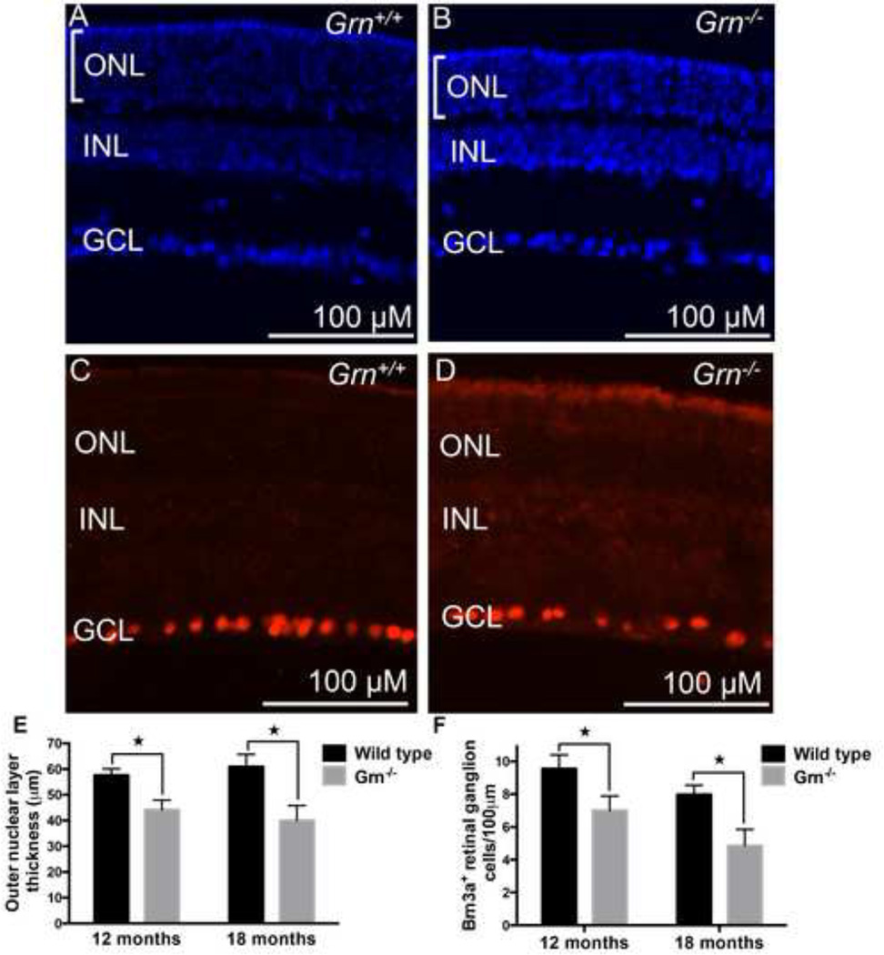
Figure 3. Loss of Progranulin leads to photoreceptor and retinal ganglion cell degeneration at 12 and 18 months of age.
(A) DAPI nuclear staining of a wild-type mouse retina and (B) Grn−/− mouse retina at 18 months age. Bars indicate the thickness of the outer nuclear layer. (C) Immunohistochemistry of wild-type mouse retina for the retinal ganglion cell maker Brn3a, and (D) Grn−/− mouse retina for Brn3a at 18 months. (E) Quantification of outer nuclear thickness was carried out for multiple retinas. The overall thickness of the outer nuclear later is shown for 12 and 18 month wild-type and Grn−/− mouse retinas. (F) Quantification of Brn3a+ cells/100 µm for wild type and Grn−/− mouse retinas at 12 and 18 months. *P<0.05, n=at least 3 mice per group. Error bars = s.d. Measurements were taken in the central regions of the retina.

