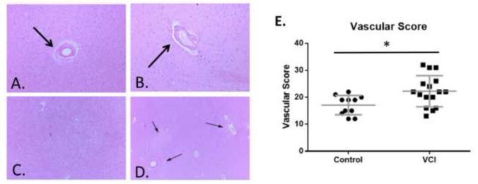
Figure 2. Histopathological characterization of control and VCI brain tissue.
Examples of vascular thickening (arrow, A.), dilated perivascular space (arrow B.), diffuse white matter rarefaction (C.), and multifocal rarefaction (arrows, D.) in aged human brain tissue. E. Comprehensive histopathological score of cerebrovascular pathology in control and VCI brain. n = 11 for control and n=16 separate brains for VCI group. Significance was determined by a Mann-Whitney non-parametric test. * indicates p<.05.

