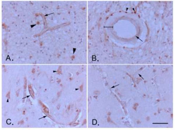
Figure 4. Expression of soluble epoxide hydrolase (sEH) in the aged human brain.
A. Endothelial staining of a white matter microvessel, likely capillary (arrow; approximate diameter 10 μm; see scale bar in D.). Other immune-positive cells in the image include astrocytes (arrowheads). B. Immunohistochemical staining of a human brain surface pial arteriole (approximate diameter of 150 μm; see scale bar in D.) showing positive staining in perivascular adventitial cells (arrowheads, likely inflammatory cells), but only weak staining in endothelial cells (arrow; compare to microvascular staining in A., which is taken from the same brain section), and no staining in tunica media vascular smooth muscle (bracketed). C. and D. Microvascular endothelial staining shows is intensified near a cortical microinfarct (arrows in C.) compared to staining in control microvessels in the same brain section away from the microinfarct (arrows in D.). Arrowheads in C. point to neurons, staining of which does not intensify near lesion. Scale bar represents 50μm. All images are at the same magnification.

