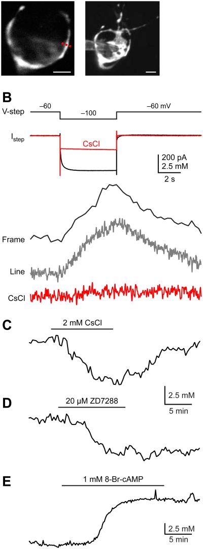
Figure 2.
Presynaptic HCN channels control resting Na+ concentration. (A) Left panel: A single optical section of a calyx of Held using two-photon microscopy; Right panel: Maximum intensity montage of the calyx. Scale bars are 5 μm for both panels. (B) A voltage step to from −60 mV to −100 mV evoked an inward current. Meanwhile, a [Na+] increase was visualized with SBFI under either frame-scan or line-scan modes. Both the inward current and Na transients were blocked by 2 mM CsCl. TTX was added to the ACSF to block voltage-gated Na+ channels. (C-D) After dialysis with SBFI and Alexa 594, the recording pipettes were subsequently detached from calyces and the Na+ signals were measured. At resting potentials, application of CsCl (C) or ZD7288 (D) reduced intracellular Na+ concentrations. (E) 8-Br-cAMP increased intracellular resting Na+ concentration. Note the difference in time scales in B and C-E.

