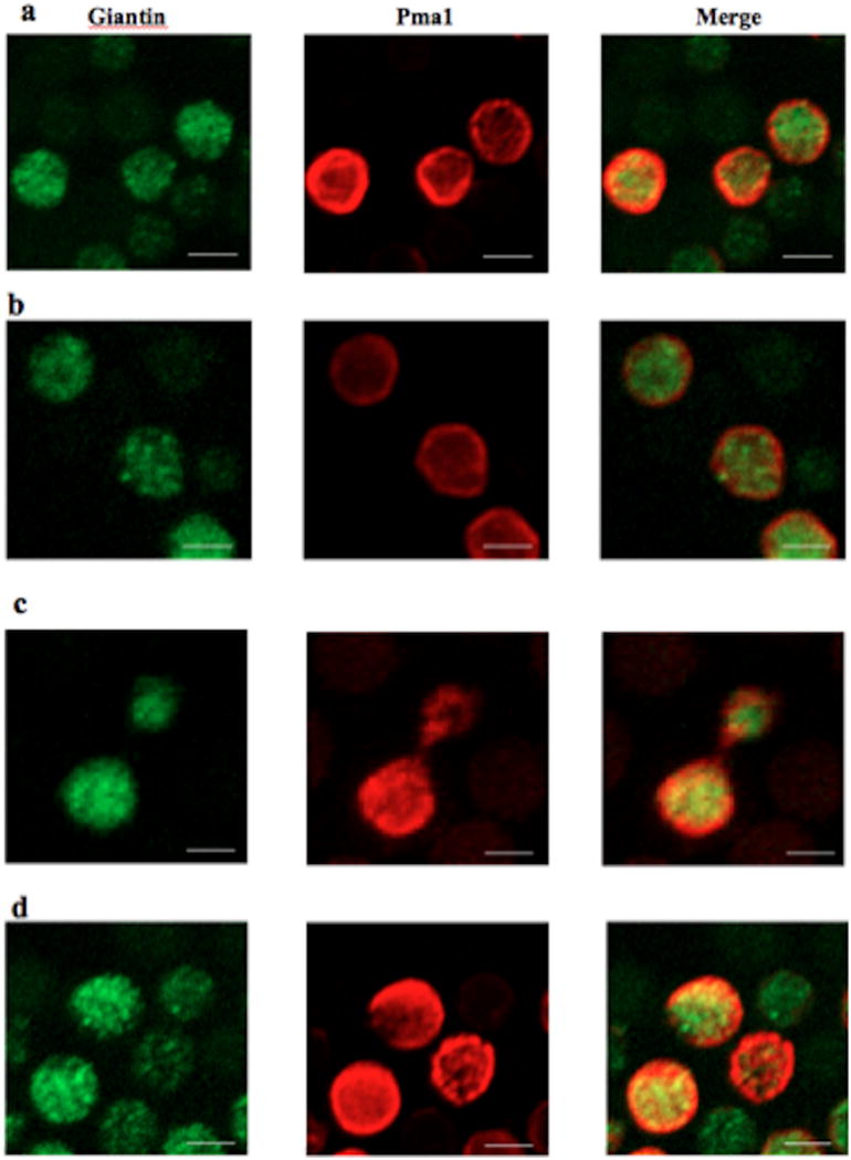
Figure 1.

Confocal imaging showing the localization of Pma1 (red) with regard to Giantin, a marker of the Golgi apparatus (green). (A) ISC1-PMA1::HA (WT), pH=4; (B) Δisc1-PMA1::HA, pH=4; (C) ISC1-PMA1::HA (WT), pH=7; (D) Δisc1-PMA1::HA, pH=7. All images were acquired after 2 hours of culture growth followed by fixing and staining. Scale bar= 10 µm.
