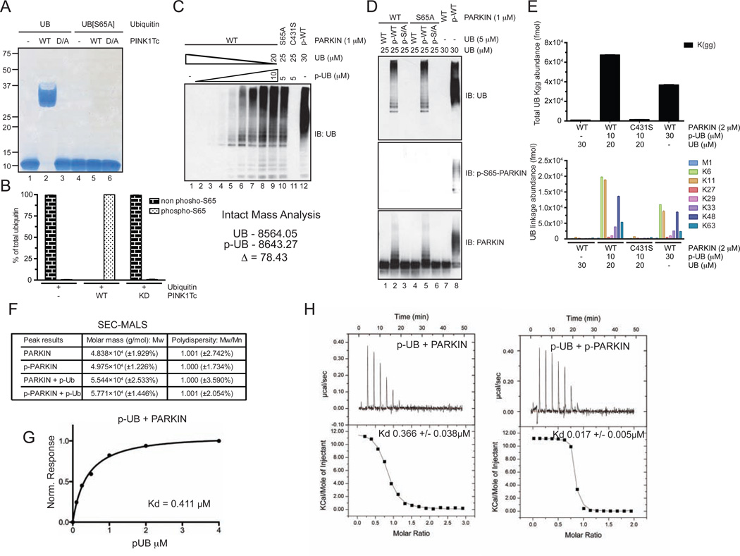
Figure 5. UB chain synthesis by and biophysical analysis of p-UB-PARKIN complexes in vitro.
(A) UB or UBS65A was incubated with PINK1Tc and reaction products analyzed using Phos-Tag gel electrophoresis.
(B) Stoichiometry of S65 phosphorylation was determined by AQUA. The intact mass of purified p-UB is also shown.
(C,D) p-UB but not p-UBS65A activates unphosphorylated PARKIN UB ligase activity in vitro. The concentrations of p-UB used in panel C were 0, 0.1, 0.25, 0.5, 1, 2, 3.5, 5, 10 µM and PARKIN was 1 µM. The total UB concentration was 30 µM.
(E) p-UB promotes the assembly of K6, K11, K48, and K63 chains by unphosphorylated PARKIN.
(F) Inactive and active forms of PARKIN are monomeric and PARKIN binds a single molecule of p-UB. Mass determination was performed by SEC-MALS.
(G) Binding of p-UB (99% phosphorylated) to PARKIN was measured using Bio-layer interferometry.
(H) Isothermal calorimetry of the indicated protein pairs.
See also Figure S5.

