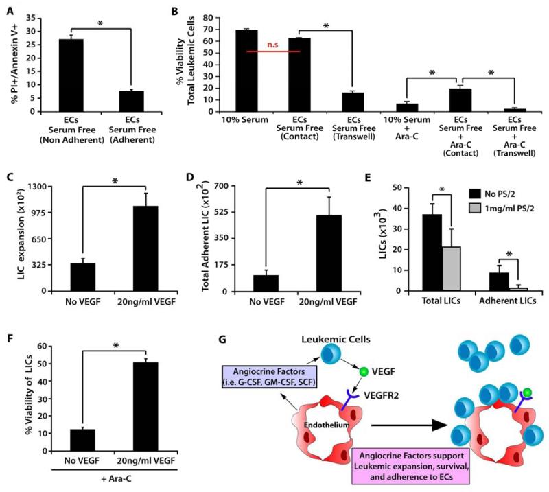
Figure 3. VEGF-A activated ECs stimulate proliferation and adhesion of leukemic cells and confer chemotherapeutic protection.
A) Annexin/PI staining to determine the number of non-viable cells present among the adherent and non-adherent populations of leukemic cells co-cultured on VeraVec. Note the increase in the percentage of live cells in the EC-adherent population. B) To determine if VeraVec might also provide a chemoprotective environment for adherent leukemic cells, leukemic cells were cultured with no feeder layer in 10% serumor co-cultured with VeraVec in either direct contact separated by transwells in serum-free conditions. These conditions were analyzed alone and with the chemotherapeutic agent Ara-C. Of those with Ara-C, only leukemic cells with direct contact to VeraVec were protected. C-E) 20 ng of VEGF-A was used to pre-stimulate VeraVec for 48 hours prior to coculture with LICs fostering a rapid increase in expansion of both the C) total and D) adherent populations of LICs after 7 days in coculture. E) VLA-4 neutralizing antibodies were added to the coculture conditions and total LIC expansion and the number of adherent LICs were analyzed after 7 days of coculture. F) 20 ng of VEGF-A increased the cell viability of LICs co-cultured on VeraVec in the presence of Ara-C, enhancing the chemoprotective properties of the VeraVec. Data was generated with 6 individual MLL-AF9 clones and each clone tested in technical triplicates in 3 independent experiments. All data represents mean + s.d. (*P<0.05, n.s.=not significant).

