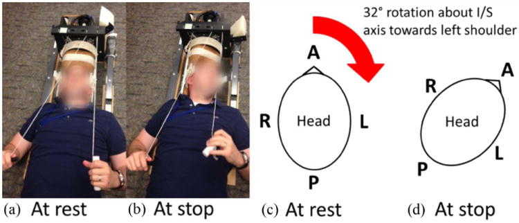
Figure 1.

Photograph of the head rotation device and schematic of head at rest and stop positions :A mild angular acceleration was generated using an MRI-compatible head rotation device.(a,c) At rest, the subject lies in the supine position with his or her head facing straight ahead. The subject initiates the motion by releasing a latch, which allows for free rotation about the inferior(I)/superior(S) axis towards his or her left shoulder. (b,d) After a 32° rotation, the device encounters a stop, which generates a mild angular acceleration. The subject is instructed to pause for 1-2 seconds at the stop. The subject then rotates his or her head back to the rest position, and the motion is repeated when the subject is ready.
