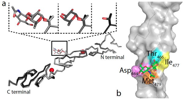
Figure 2. Structure of O-fucosylated variants of human Notch1 EGF repeats 11–13.

(a) Superimposed X-ray structures of the unmodified human Notch1 EGF11–13 (PDB ID code 2VJ3) and the monosaccharide and disaccharide structures with the subsequent additions to the O-fucose site (Thr466) region highlighted. Calcium ions are shown in red. (b) Details of human Notch1 EGF12 X-ray structure highlighting contacts between the C6 methyl group of the O-fucose with Ile477 (yellow) and Met479 (orange), the C6 methoxy group of GlcNAc with Asp464 (violet), and the N-acetyl group of GlcNAc with Met479 (orange). Thr466 is highlighted in cyan. Reproduced with permission from [56].
