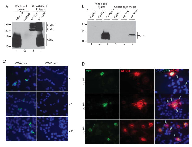
Figure 4. Agnoprotein is taken up by glial cells.
A. TC620 cells were transduced with either a null adenovirus or adenovirus encoding agnoprotein. Growth medium was collected in parallel to whole cell protein lysates at 48 h post-transduction. Agnoprotein was immunoprecipitated from growth medium and analyzed by Western blot (lanes 3 and 4) as described in Fig. 1C. Whole cell protein lysates were also loaded as positive and negative controls of expression (lanes 1 and 2, respectively). B. Western blot analysis of whole cell extracts from cells expressing agnoprotein (lanes 1–3) or from cells treated with conditioned medium obtained from control, adeno-null, and adeno-agno transduced cells. C. TC620 cells were seeded in two-well chamber slides and treated with growth medium from agnoprotein-expressing cells or control cells. At 3, 9, and 24 h post-treatment, cells were washed 3X with PBS and fixed with cold acetone/methanol (1/1). Immunocytochemistry was performed to detect agnoprotein as described in materials and methods. D. SVGA cells were transfected/infected with Mad-1 JCV and cells were fixed on 2-well chamber slides at 10, 20, and 30 days post-infection. Expression and co-localization of VP1 and agnoprotein were analyzed by immunocytochemistry. Nuclei were also labeled with DAPI (blue) and merged with the agno (red) and VP1 (green). Arrows point cells positive for agno (red) and negative for VP1 (green) fluorescein.

