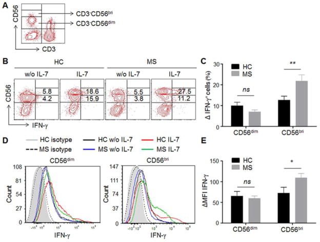
Fig. 2. IL-7 stimulation leads to a higher increase of IFN-γ production in NK cells of MS patients.
(A) Gating strategy for NK subpopulations. NK cells in PBMCs are analyzed by flow cytometry and gated based on surface markers CD3 and CD56, which express CD3−CD56dim or CD3−CD56bright. (B) Representative IFN-γ expressing NK cells from one MS patient and one HC with or without IL-7 stimulation are shown. PBMCs from MS patients and HCs were cultured with or without IL-7 for 72 hour. Intracellular staining of IFN-γ gating on CD3−CD56dim and CD3−CD56bright NK cells were analyzed by flow cytometry. (C) Increase in the percentage of IFN-γ-expressing CD56dim and CD56bright cells was calculated as the difference between unstimulated and IL-7 stimulation. (D) Representative intracellular IFN-γ intensity on NK cells from one MS patient and one HC is shown. (E) The changes in IFN-γ MFI on CD56dim and CD56bri cells were calculated as the difference between unstimulated and IL-7 stimulation. Data from 10 HCs and 10 MS are summarized. 5 relapsing remitting patients, 3 secondary progressive and 2 primary progressive patients were included in this study. HC were relatives of MS patients. *p < 0.05; **p < 0.01. ns, not significant. MFI, mean fluorescence intensity. CD56bri indicates CD56bright.

