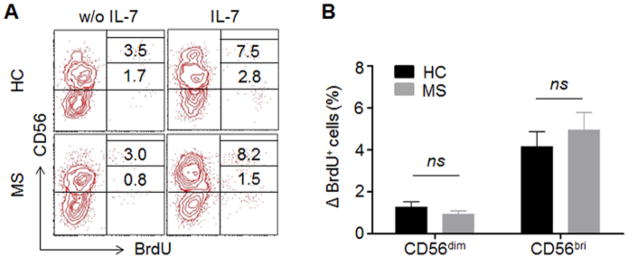
Fig. 4. No differences in IL-7-induced proliferation.

PBMCs from MS patients and HCs were cultured with cytokine IL-7 or left unstimulated without IL-7 for 72 hours. Proliferation of NK cells were analyzed by BrdU incorporation assay. (A) Representative fluorescence-activated cell sorting (FACS) plot showing proliferating NK cells (BrdU+) from one MS patient and one HC. (B) The changes on the percentage of BrdU+CD56dimand BrdU+CD56bright cells were calculated between unstimulated and IL-7 stimulation. n = 10 per group. 5 relapsing remitting patients, 3 secondary progressive and 2 primary progressive patients were included in this study. HC were relatives of MS patients. CD56bri indicates CD56bright. ns, not significant.
