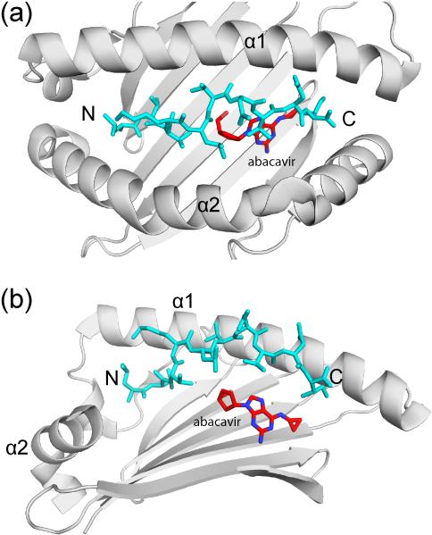
Figure 3. Induction of T cell mediated immunopathology by binding of a small molecule drug in the MHC class I groove.
Crystal structure of a HLA-B57:01 – peptide complex with bound abacavir, an anti-retroviral drug (PDB ID 3VRJ). Top view (a) and side view (b) of the peptide binding groove, showing how abacavir (red) is bound underneath the peptide (blue). In the side view, the HLA-B α2 helix has been removed to aid visualization of abacavir. The α1 and α2 helices of the HLA-B heavy chain are indicated as well as the N and C terminus of the bound peptide (N and C, respectively).

