
Figure 2. Schematic structure(A), restriction enzyme digestion and PCR identification(B) of recombinant plasmid pBacPAK8-CTB-Aβ42.
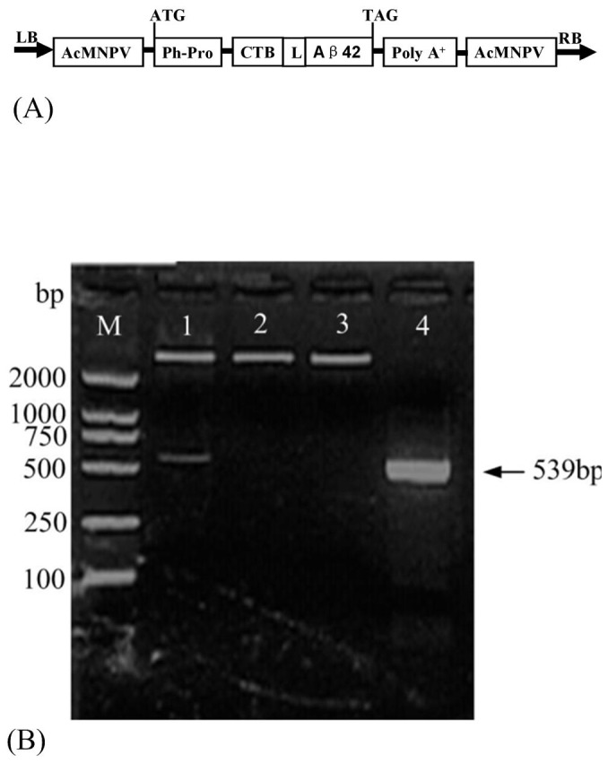
(A) LB, left border; AcMNPV, Autographa Californica Nuclear Polyhedrosis Virus; Ph-Pro, AcMNPV polyhedrin promoter; L, linker peptide (GPGP); poly A+, polyadenylation signal; RB, right border. (B) M, DNA marker DL 2000 (TakaRa); lane 1, recombinant plasmid digested with BamHI and Xho I; lane 2, recombinant plasmid digested with BamHI; lane 3, recombinant plasmid digested with Xho I; lane 4, PCR amplification of recombinant plasmid.