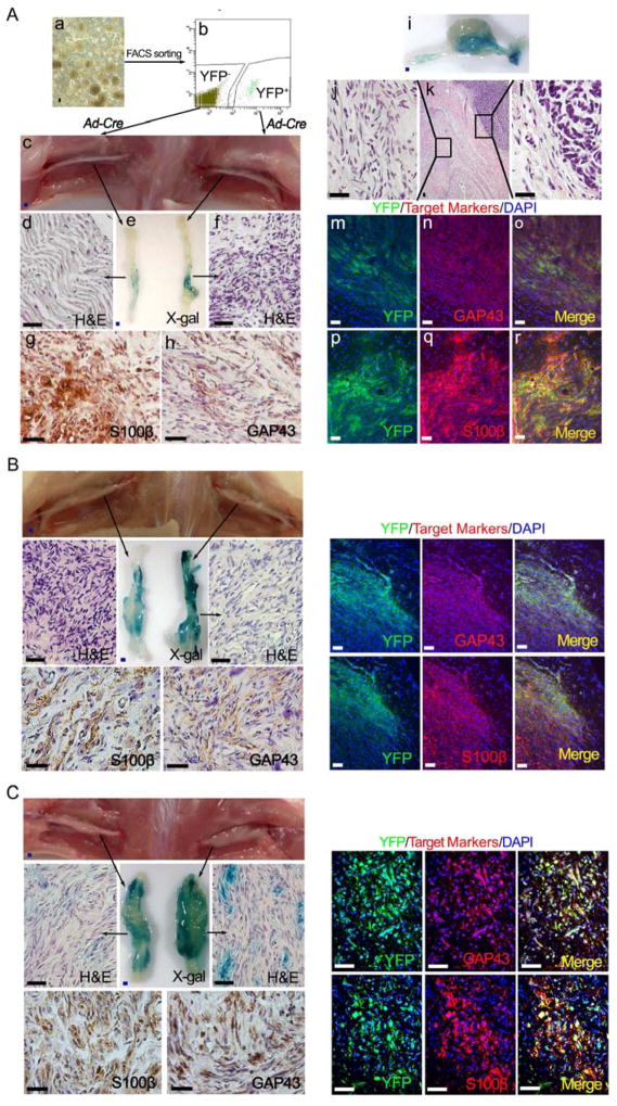
Figure 2. The cells of origin for para-spinal plexiform neurofibroma are inside the embryonic PLP+ DNSCs.
(A) FACS was performed to obtain the YFP+ and YFP− DNSCs from E13.5 PlpCre-ERT;Nf1flox/flox;R26R-LacZ-YFP embryo, whose mother was administered with tamoxifen at E11.5 (a and b); Both YFP+ and YFP− DNSCs were infected with Ad-Cre to ablate Nf1, then injected to right and left sciatic nerves of nude mice respectively (c); X-gal and H&E staining were perform on left and right sciatic nerve (d – f); Sections of right sciatic nerve were stained with antibody against S100β (g) and GAP43 (h); A fraction of these sciatic plexiform neurofibromas spontaneously transformed into MPNST, exhibiting both benign and malignant histologic characters (i – l); Immunofluorescence staining of tumor on right sciatic nerve shows expression of YFP as well as GAP43 and S100β Schwann cell markers (m – r).
(B and C) A similar strategy to that in panel A was used for YFP+ and YFP− DNSCs derived from E13.5 embryos with genotype Krox20-Cre;Nf1flox/flox;R26R-LacZ-YFP (B) and Dhh-Cre;Nf1flox/flox;R26R-LacZ-YFP (C).
Scale bars: 500 μm for blue scale bars, 50 μm for white and black scale bars.

