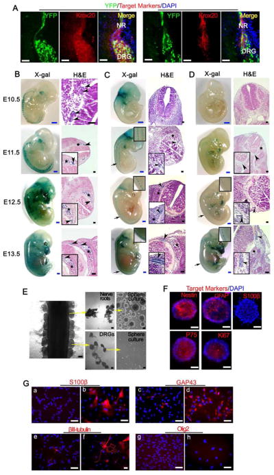
Figure 3. Cells of origin for para-spinal plexiform neurofibromas are inside the embryonic PLP+ nerve root cells.
(A) Representative frozen sections from E13.5 PlpCre-ERT;Nf1flox/flox;R26R-LacZ-YFP embryo, whose mother was administered with tamoxifen at E11.5, were stained for YFP, Krox20, DHH and DAPI. NR=nerve root, DRG=dorsal root ganglia.
(B, C, and D) X-gal and H&E staining were performed on embryos with genotype PlpCre-ERT;R26R-LacZ (B, mother mouse was administered with 4-hydroxytamoxifen at E9.5), Krox20-Cre;R26R-LacZ (C) and Dhh-Cre;R26R-LacZ (D) from E10.5 to E13.5. Arrow = area of enlarged view, arrow head = nerve roots, star mark = DRG.
(E) DRG and dorsal nerve root were separated from E13.5 embryos and cultured in neurosphere culture conditions.
(F) Nerve root derived neurospheres immunostained for Nestin, GFAP, S100β, P75, Ki67 and DAPI.
(G) Immunostaining of nerve root derived neurosphere cells cultured in neurosphere medium (a, c, e, and g) or differentiation medium (b, d, f, and h) with antibodies against S100β, GAP43, βIII-tubulin and Olig2.
Scale bars: 500 μm for blue scale bars, 50 μm for white and black scale bars.
See also Figure S2.

