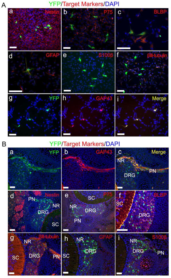
Figure 4. Phenotypic analysis identifies the PLP+ cells of origin as embryonic Schwann cell precursors.
(A) DNSCs obtained from E13.5 PlpCre-ERT;Nf1flox/flox;R26R-YFP embryos, whose mother was administered with tamoxifen at E11.5, were stained for YFP, DAPI and lineage markers: Nestin (a), P75 (b), BLBP (c), GFAP(d), S100β (e), βIII-tubulin (f) and GAP43 (g – i).
(B) Formalin fixed paraffin embedded sections from E13.5 PlpCre-ERT;Nf1flox/flox;R26R-YFP embryos, whose mother was administered with tamoxifen at E11.5, were stained for YFP, DAPI and lineage markers: GAP43 (a– c), Nestin (d), P75 (e), BLBP (f), βIII-tubulin (g), GFAP (h) and S100β (i). DRG=dorsal root ganglia, NR= nerve root, PN=peripheral nerve, SC=spinal cord.
Scale bars: 50 μm for all.
See also Figure S3.

