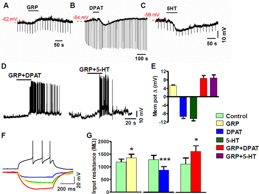
Figure 6. Co-Activation of 5-HT1A and GRPR Increased the Excitability of GRPR+ Neurons.
(A-C) Representative traces show membrane depolarization by GRP (A) and hyperpolarizing response induced by DPAT (B) and 5-HT (C) in current-clamped GRPR+ neurons.
(D) Representative traces showing that a co-application of GRP and DPAT or 5-HT evoked membrane depolarization and AP firing in current-clamped GRPR+ neurons. AP firing evoked by GRP+DPAT and GRP+5-HT ranged 0.167 to 3.5 Hz (n = 6) and 0.03 to 6 Hz (n = 17), respectively.
(E) Quantified data of A-D. GRP depolarized GRPR+ neurons by 5.3 ± 0.3 mV (n = 8). DPAT (7.5 µM, blue) and 5-HT (40 µM, dark green) hyperpolarized GRPR+ neurons by 9.4 ± 0.9 mV (n = 19) and 10.4 ± 1 mV (n =13), respectively. In contrast, co-application of GRP+DPAT depolarized GRPR+ neurons by 8.7 ± 1.4 mV (red) (n = 15). GRP+5-HT depolarized GRPR+ neurons by 8.9 ± 1.5 mV (purple) (n = 35 ).
(F) Representative traces illustrate the effect of GRP (yellow), DPAT (blue), and GRP+DPAT (red) on membrane input resistance in current-clamped GRPR+ neurons receiving negative current injections (−20 pA). Control (green): extracellular buffer only. For all recordings, neuronal health was verified by observing action potentials in response to positive current injection under control conditions (black).
(G) GRP (yellow) increased the membrane input resistance from 1,185 ± 124 MΩ to 1,350 ± 150 MΩ (n = 8), DPAT decreased the input resistance from 1,311 ± 168 MΩ to 894 ± 137 MΩ (n = 19) and GRP + DPAT increased the input resistance from 1,116 ± 247 MΩ to 1,616 ± 226 MΩ (n = 15), *p < 0.05, **p < 0.01, ***p < 0.001, paired t-test.

