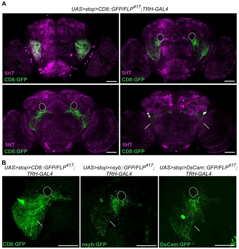
Figure 3. Anatomical characterization of the aggression-modulating 5HT-PLP neurons.
A. Arborization patterns of the PLP neurons visualized by membrane-bound CD8::GFP (green) relative to anti-5HT labeled neuropil regions of the brain (magenta). These are displayed in frontal z-projections of an image stack through the ventrolateral protocerebrum and antennal lobes (top left), the ellipsoid body of the central complex and the peduncles of the mushroom bodies (top right), the fan-shaped body of the central complex (bottom left) and a posterior view of the brain where the PLP cell bodies and their axons are located (bottom right). Scale bar represents 50 μm. Short arrows point to cell bodies, long arrows to axons of the PLP neurons. A dotted line outlines the peduncles of the mushroom bodies that are not stained by anti-5HT antibodies.
B. Polarity of the serotonergic PLP neurons. (Left) The total arborization field of the PLP neurons visualized using membrane-bound CD8::GFP. (Center) The putative presynaptic terminals of the PLP neurons revealed using the presynaptic marker nsyb::GFP. (Right) The putative dendritic arbors of the PLP neurons visualized by expression of the postsynaptic marker DsCam:GFP. Full z-stack frontal projections are shown, scale bar represents 50 μm.

