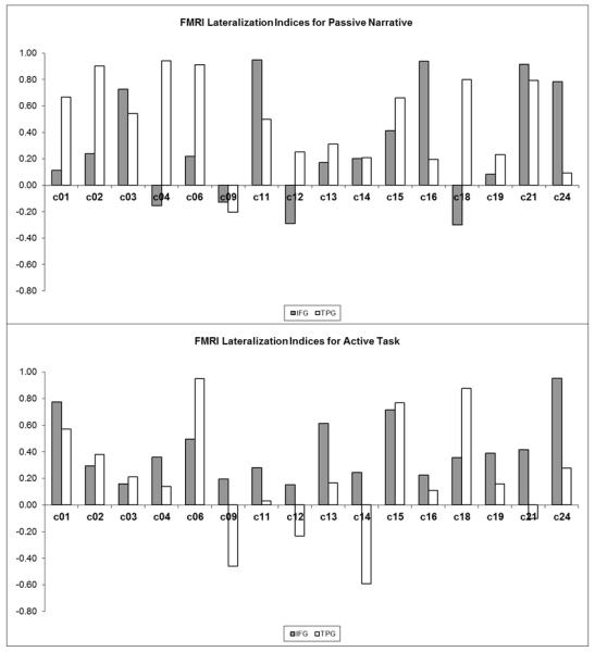
Figure 6.

Control cohort lateralization indices (LI) for the inferior frontal gyrus (IFG) and temporoparietal (TPG) language regions, using activate language fMRI (top panel) and passive language fMRI (bottom panel). Positive LI values indicate left-lateralization, negative LI indicates right-lateralization, and LI values of magnitude 0.1 or less indicate bilateral activation.
