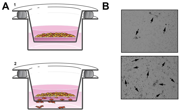
Figure 2. Boyden chamber assay.
(A) Graphical depiction of an invasion assay. 1. HNSCC cells seeded on BME in the upper chamber. 2. Cells invade toward a chemoattractant. (B) Photographs show HNSCC cells (highlighted by arrows) that have penetrated the BME. The upper image shows less invasion than the lower image.

