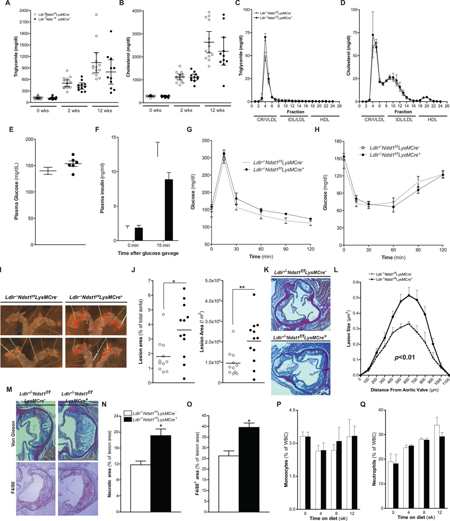
Figure 2. Macrophage NDST1 Inactivation Increases Atherogenesis and Lesion Macrophage Content.
Plasma cholesterol and triglycerides levels from female Ldlr−/−Ndst1f/f and Ldlr−/−Ndst1f/fLysMCre+ mice at different times after commencing HFC diet feeding (A and B). FPLC analysis of plasma after 12-weeks HFC diet feeding (n = 6) (C and D). Fasting plasma glucose and insulin levels before and after an oral glucose gavage (n=5–7) (E and F). Glucose tolerance (G) and insulin tolerance (G) tests were performed after 11-weeks HFC diet feeding (n=5–7). En face analysis of atherosclerosis and quantification of Sudan IV positive area (E and F). Staining and quantification of atherosclerotic lesion size in the aortic sinus (n = 5–7) (G and H). Atherosclerotic lesions stained with Van Gieson for collagen or F4/80 for macrophages (I). Quantification of necrotic core size (J) and F4/80 positive area (K) in lesions after 12-weeks HFC diet feeding. Monocyte(L) and Neutrophil (L) counts in blood during HFC diet feeding. Shown are mean values ± SEM, *p<0.05 and **p<0.01.

