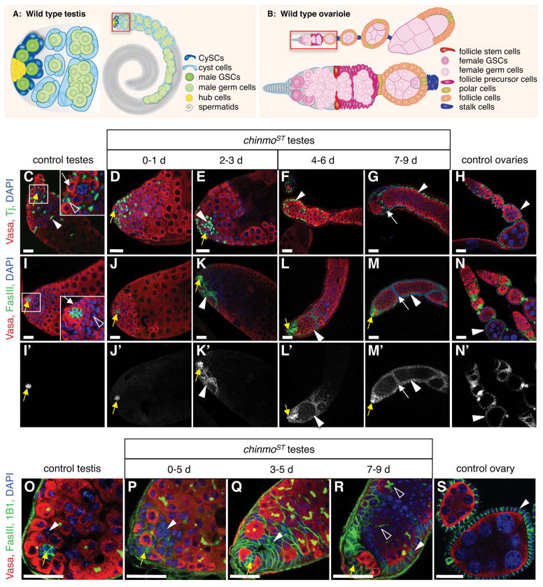
Figure 1. Reduction of Chinmo causes somatic cells in adult testes to be gradually replaced by cells resembling ovarian follicle cells.
(A) Illustration of a wild-type Drosophila testis (right) with the apex magnified (left). Germline stem cells (GSCs, dark green) and somatic cyst stem cells (CySCs, dark blue) adhere to the hub (yellow). GSCs produce differentiating male germ cells (spermatogonia and spermatocytes, green) that are displaced from the hub and form elongated spermatids (grey) and mature sperm (not shown). Approximately two CySCs flank each GSC; CySCs produce squamous, quiescent cyst cells (light blue), which encase differentiating germ cells. (B) Illustration of a wild-type Drosophila ovariole (top) comprised of a germarium (magnified, bottom) followed by a series of developing egg chambers. In the germarium, anterior niche cells cap cells (grey) support GSCs (dark pink), which produce differentiating female germ cells (light pink). Two somatic follicle stem cells (red), located near the middle of the germarium, produce follicle precursor cells (magenta), which differentiate into follicle cells (orange), stalk cells (purple), and polar cells (yellow). Each egg chamber contains 16 germ cells surrounded by a monolayer of columnar epithelial follicle cells. Polar cells are located at each end; egg chambers are linked by chains of stalk cells. (C–S) Immunofluorescence detection in adult testes and ovaries of Tj (C–H, green) to visualize somatic cell nuclei, or FasIII (I–S, green) to highlight the hub in all testes (yellow arrows), and somatic cell membranes in ovaries and chinmo mutant testes (arrowheads). Panels I′-N′ show the FasIII signal alone. Vasa (red) marks germ cells and DAPI (blue) marks nuclei in all panels. In control testes (C, I, O), somatic CySC lineage cells (arrowheads) are squamous and interspersed among germ cells. Insets (C, I) show GSCs (white arrows) and CySCs (open arrowheads) surrounding the hub. In chinmoST testes (D–G, J–M, P–R), a distinct phenotype develops over time. Testes from young mutant males (D–E, J–K, P–Q) resemble those from controls except that most (~77%, n = 61) contain aggregates of 8 or more somatic cells (arrowheads); these always appear near the hub (yellow arrows). As flies age (F–G, L–M, R), aggregates expand beyond the testis apex and become columnar and peripheral (arrowheads) in 82% of testes (n = 545), forming FasIII-positive “follicle-like cells” that resemble somatic follicle cells (arrowheads) in control ovaries (H, N, S). Follicle-like cells occasionally invaginate (G, M, white arrows) to envelop groups of germ cells. 1B1 (O–R, green) marks fusomes; branching fusomes in older germ cells in chinmoST testes indicate spermatogonial arrest (R, open arrowheads). Scale bars = 20 μm. See also Figure S1.

