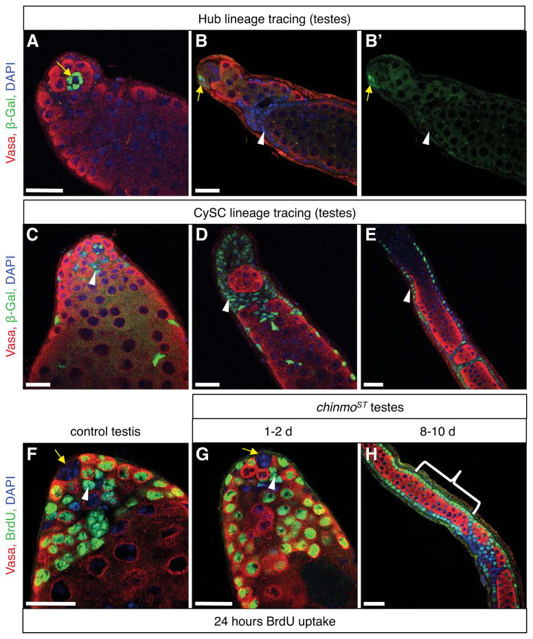
Figure 6. Follicle-like cells come from the cyst stem cell lineage but not from hub cells.
(A–E) Immunofluorescence detection of β-gal (green), which permanently marks either hub cells alone (A–B) or CySC lineage and hub cells (C–E) in chinmoST testes. Somatic cell aggregates and follicle-like cells (B, D–E, arrowheads) are derived from CySC lineage cells (C, arrowhead) but not from hub cells (A–B, arrows) because they express β-gal in testes with marked CySC lineage cells but not in testes with only marked hubs. (F–H) Immunofluorescence detection of the thymidine analog bromodeoxyuridine (BrdU, green). Adult males were fed BrdU for 24 hr prior to dissection to label all cells that traversed S-phase during this time. In control (F) and young chinmoST (G) testes, BrdU is not found in any hub cells (arrows), but many germ cells (red) and CySCs (arrowheads) are BrdU+. BrdU is also found in most follicle-like cells in older chinmoST testes (H, bracket). In all panels, DAPI marks nuclei (blue) and Vasa marks germ cells (red). Scale bars = 20 μm. See also Table S4.

