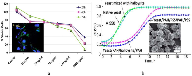
Fig. 4.

a) MTT viability test curves demonstrating the cellular viability after subjecting the MCF-7 cells to increasing concentrations of halloysite nanotubes. Insert: laser scanning confocal microscopy image of FITC-polyelectrolyte coated halloysite nanotubes (green) located near cellular nuclei (blue) of cultured MCF-7 cells. Reproduced with permission from [47]; b) growth curves of native yeast cells, yeast mixed with halloysite nanotubes, yeast coated with PAH/PSS/PAH/PSS and yeast coated with PAH/HNTs/PAH/PSS. Insert: SEM image of yeast cells coated with halloysite-doped polymer shells. Reprinted with permission from [45]. Copyright (2013) The Royal Society of Chemistry.
