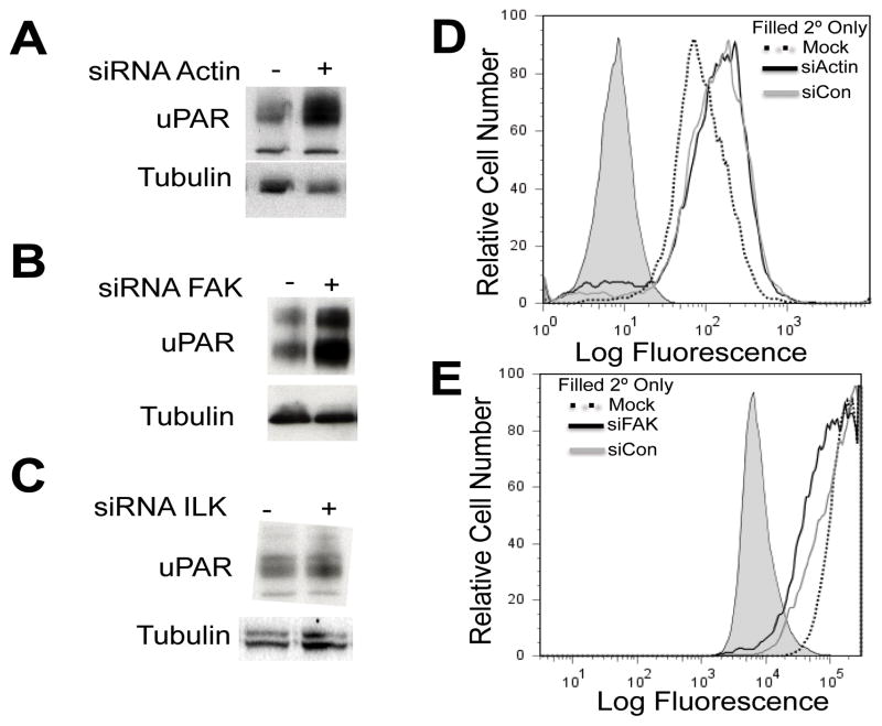
Fig. 3.
(A) DU145 cells were transfected with siRNA targeting (A) actin for 96 h (B) FAK for 72 h and (C) ILK for 72 h. Immunoblot analysis using whole cell lysates was used to detect uPAR expression for each experiment. DU145 cells were transfected with siRNA targeting (D) actin for 96 h and (E) FAK for 72 h and cell surface expression of uPAR was detected by flow cytometry using the mAB 3936. Independent experiments were performed three times.

