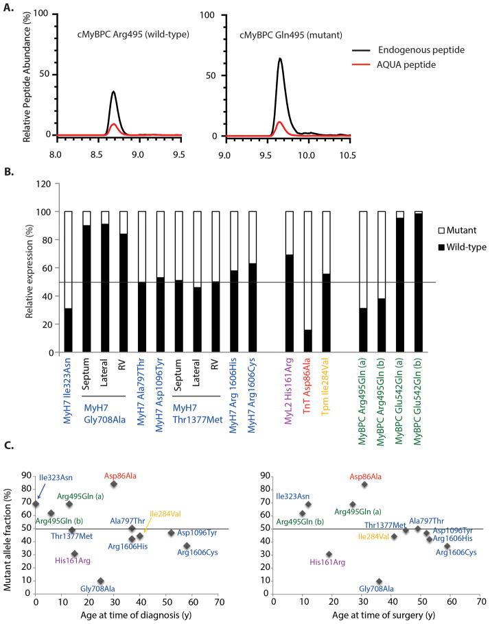
Figure 4.
Stoichiometry of wild-type and mutant sarcomere protein in human HCM determined by absolute quantification of abundance (AQUA). (A) Representative chromatogram from HCM sample MYBPC3 Arg495Gln (a) showing peptide abundance of wild-type and mutant peptides relative to their corresponding AQUA peptides. (B) Graph summary of relative peptide abundance of wild-type and mutant peptides from individual samples. The abundance of each peptide(s) (fmol) was calculated, summed in cases of missed cleavages or post-translational modifications (Supplementary Table 4), and expressed as a relative percentage of the total (abundance of wild-type + mutant peptides for each sample). Three regions (septum, LV lateral wall, and right ventricle) were analyzed in 2 samples with MYH7 mutations obtained from explanted hearts at the time of transplantation. For both B and C, color coding is used to designate genes harboring mutations: MYH7 (blue), MYBPC3 (green), TNNT2 (red), TPM1 (yellow), MYL2 (purple). (C) Relationship of patient age at time of surgery (left panel) and age at time of diagnosis of HCM (right panel) to mutant allele fraction. There was a significantly greater degree of allelic imbalance (in either direction) in patients who had surgery ≤ 40 y/o, compared to those > 40 y/o (P<0.01) and in patients who were diagnosed ≤ 30 y/o, compared to those diagnosed > 30 y/o (P<0.05).

