
Figure 1.
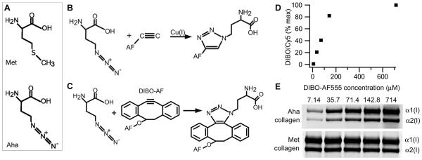
Procollagen labeling with azidohomoalanine (Aha). (A) Aha and methionine (Met) structures. (B) Cu-catalyzed and (C) Cu-free conjugation of Aha with Alexa Fluor (AF) fluorescent dyes. (D) Aha conjugation efficiency with DIBO-AF555 at different concentrations of the dye calculated from AF555/Cy5 fluorescence intensity ratio in the same gel band (panel E). The conjugation efficiency at 714 uM DIBO-AF555 is assumed to be 100%. (E) Gel electrophoresis of pepsin-treated procollagen (collagen) after Aha conjugation with DIBO-AF555 and Lys conjugation with Cy5. Aha-labeled collagen is revealed by fluorescence scanning at 532 nm excitation and 570±10 nm emission wavelength (AF555) while total collagen (Met collagen) is revealed by scanning of the same gel bands at 635 nm excitation and ≥ 665 nm emission (Cy5).