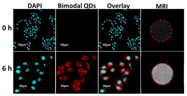
Figure 6.

The confocal fluorescence and MR images of HeLa cells before and 6 h after incubation with GZCIS/ZnS@BSA at the Gd concentration of 0.1 mM. The cell nuclei were stained with DAPI. Here involved GZCIS/ZnS QDs showed PL emission at 625 nm and were prepared with Gd/Cu and Zn/Cu ratio fixed at 4/1 and 1/2, respectively.
