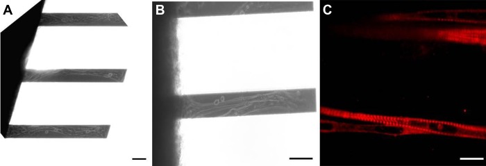
Fig. 1.

Images of adult-derived skeletal muscle myotubes. Phase-contrast images are shown of myotubes on silicon cantilevers at ×10 (A) and ×20 (B) magnification. C: immunostaining of cultures using an antibody against myosin heavy chain (MHC) with all classes (A4.1025, DSHB) indicating highly striated and mature muscle fibers. Scale bar = 100 μm.
