
Fig. 2.
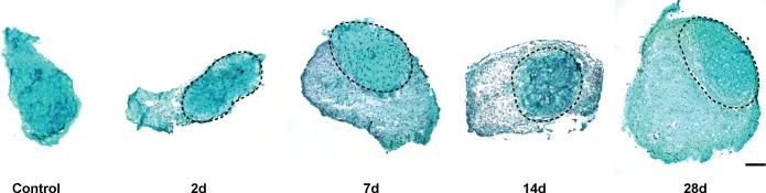
Representative cross sections of plantaris tendons from control and synergist ablation groups. Sections stained with fast green and hematoxylin. The original tendon in all groups subjected to synergist ablation is circled in a dashed line, and the neotendon matrix is located superficial to this line. Scale bar for all panels = 100 μm.