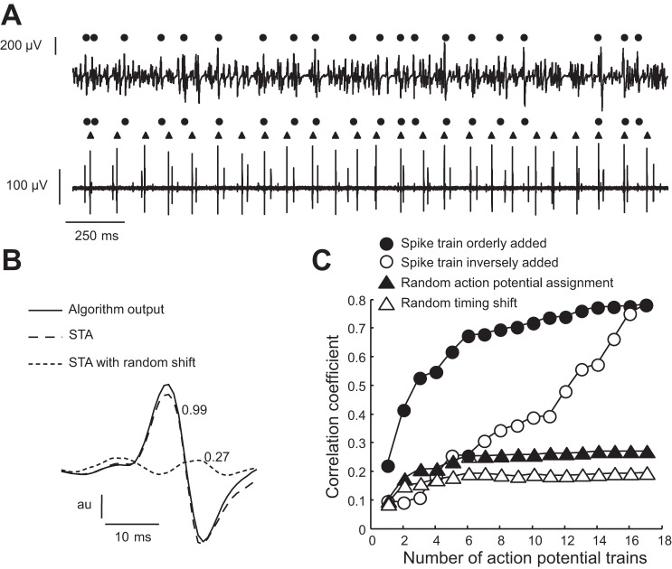
Fig. 9.
Validation of a new “decomposition” algorithm based on the criteria described by Hu et al. (57). A: surface and intramuscular EMG signals were recorded concurrently during a contraction at ∼2% MVC force. The subject was provided with visual feedback on the discharge of a single motor unit from an intramuscular EMG recording (bottom trace). The motor unit with the greater amplitude action potentials was selected for the feedback signal. The algorithm described in the text identified the discharge times of several motor units in the surface EMG signal, which are indicated by the black circles. Although there was an association between the discharge of the target motor unit (triangles) and some of the discharge times detected by the algorithm, the rate of agreement was <15% even though the target motor unit had relatively high power in the surface EMG signal. B: despite the poor performance of the algorithm, spike-triggered averaging of the surface EMG signal from the discharge times identified by the new algorithm provide an action potential shape that was similar (correlation coefficient, 0.99) to that given as an output by the decomposition algorithm. This association disappeared for small perturbations (in the same range as in Ref. 57) of the discharge times, so that the correlation coefficient between the averaged potential with perturbed triggers and that provided by the algorithm decreased to only 0.27. C: progressive addition of the trains of decomposed action potentials (in order or in reverse order; filled and empty circles) produced correlation coefficients with the recorded EMG signal that increased monotonically, in contrast to the addition of perturbed discharge times (empty triangles) or random assignment of the decomposed action potentials to the trains of discharge times (black triangles). The results described in B and C have been interpreted as indicating the valid decomposition of a surface EMG signal (57), despite its inadequate performance as shown in A.

