Fig. 3.

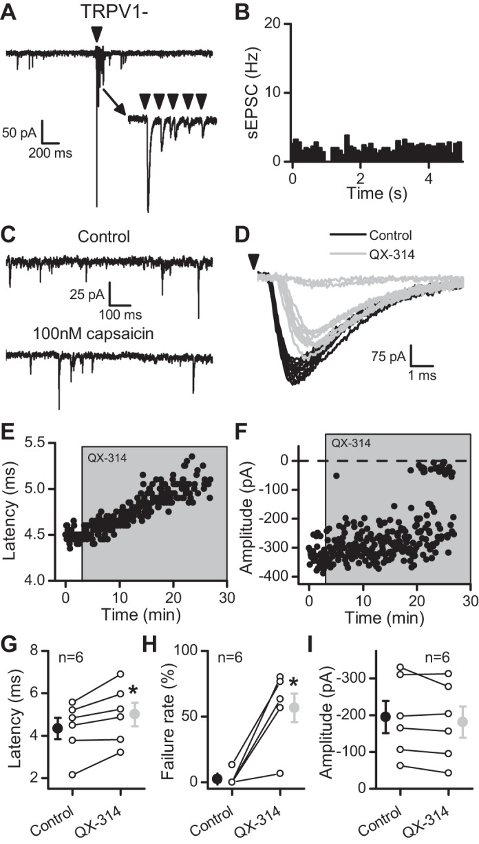
QX-314 delayed and then blocked evoked ST-EPSCs at TRPV1− afferents. In A–F, the data come from a single representative cell. A: in this single sweep, the arrow indicates stimulation of the ST (5 pulses at 50 Hz). Notice the lower rate of sEPSCs before stimulation and the lack of a transient increase in sEPSC frequency following ST stimulation compared with TRPV1+ afferents (Fig. 1). The inset demonstrates the frequency-dependent depression of the evoked response, which is similar to that seen in TRPV1+ afferents. B: TRPV1− afferents lack asynchronous release following stimulation of the ST (compare with Fig. 1B). Events were collected in 100-ms bins over 50 trials (5 min). C: this neuron lacked TRPV1 expression because capsaicin failed to increase the rate of sEPSCs (compare with Fig. 1C). D: similar to TRPV1+ afferents, 300 μM QX-314 increased the latency and caused failure of the evoked ST-EPSCs at a TRPV1− neuron. Black traces are from trials in control, whereas light gray traces are from trials in QX-314. E: the latency progressively increased following exposure to QX-314. F: the evoked ST-EPSC eventually failed in the presence of QX-314. G: the latency increased on average over 6 neurons tested (control, black; QX-314, light gray). H: summary of the failure rate of EPSC1 (control, black; QX-314, light gray). I: before failure, QX-314 did not significantly change the amplitude of ST-EPSCs (control, black; QX-314, light gray). All asterisks represent P ≤ 0.05.
