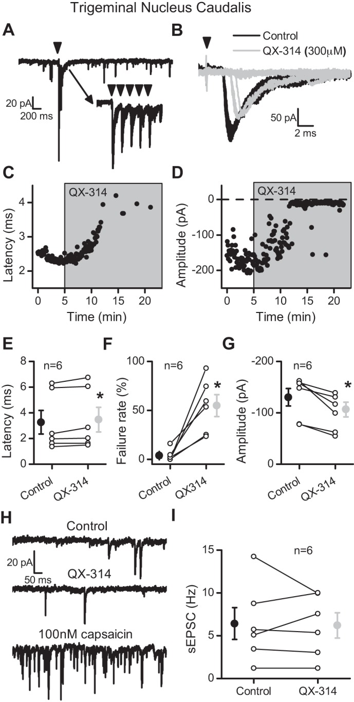
Fig. 9.

QX-314 increased the latency and then blocked evoked EPSCs at spinal trigeminal tract (spVT) afferents in caudal trigeminal (Vc) neurons. A: representative trace showing evoked EPSCs following stimulation of the trigeminal tract (5 shocks at 50 Hz). Inset traces are of individual sweeps. Arrowheads indicate the time of tract stimulation. B: QX-314 (300 μM, gray traces) prolonged the latency and caused failure compared with control (black traces). The arrowhead indicates stimulation of the trigeminal tract. C: exposure to QX-314 progressively increased the latency of the evoked spVT-EPSC. D: the amplitude of the evoked spVT-EPSC decreased slightly before resulting in failures in the presence of QX-314. E: QX-314 significantly shifted the latency of the evoked EPSCs before failures (n = 6). F: the failure rate of EPSC1 significantly increased following application of QX-314. G: the amplitudes of the evoked EPSCs significantly decreased before failure. H: representative trace of sEPSCs in control (top) and QX-314 (middle). This neuron was also sensitive to capsaicin (100 nM; bottom). I: QX-314 (light gray) did not significantly alter sEPSC rate compared with control values (black). All asterisks are for P ≤ 0.05.
