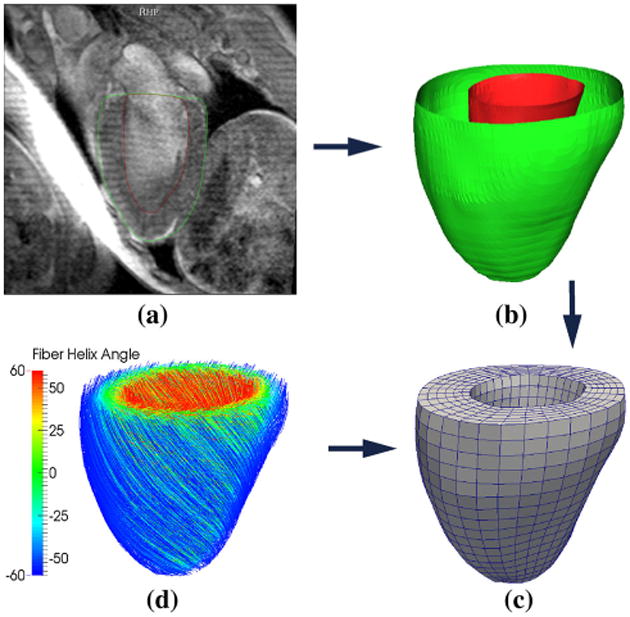
Fig. 3.

Construction of the HUMAN finite element model: (a) segmentation of the MRI, (b) reconstruction the endocardial (red) and epicardial (green), (c) construction of the finite element mesh and (d) assignment of rule-based myofiber orientation—streamlines follow fiber direction and are color coded with fiber helix angle
