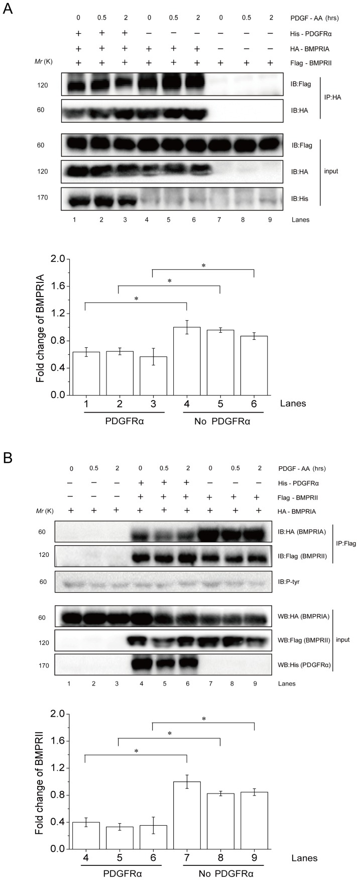
Figure 6. PDGFRα interferes with BMPRI-BMPRII interaction.
A. PDGFRα (His tagged) was co-expressed with BMPRIA (HA tagged) and BMPRII (Flag tagged) in 293T cells. The cells were treated with 25 ng/ml PDGF-AA for 0.5 or 2 h or untreated. Cell lysates were divided into two parts for analysis of BMPRIA and BMPRII expression by western blot, or immunoprecipitation of BMPRIA to test whether the level of BMPRII brought down with BMPRIA was affected by expression of PDGFRα. Right panel: quantitation data. *, p<0.05, when compared to empty vectors transfected cells. B. PDGFRα (His tagged) was co-expressed with BMPRIA (HA tagged) and BMPRII (Flag tagged) in 293T cells. The cells were treated with 25 ng/ml PDGF-AA for 0.5 or 2 h or untreated. Cell lysates were divided into two parts for analysis of BMPRIA and BMPRII expression by western blot, or immunoprecipitation of BMPRII to test whether the level of BMPRIA brought down with BMPRII was affected by expression of PDGFRα. Right panel: quantitation data. *, p<0.05, when compared to empty vectors transfected cells.

