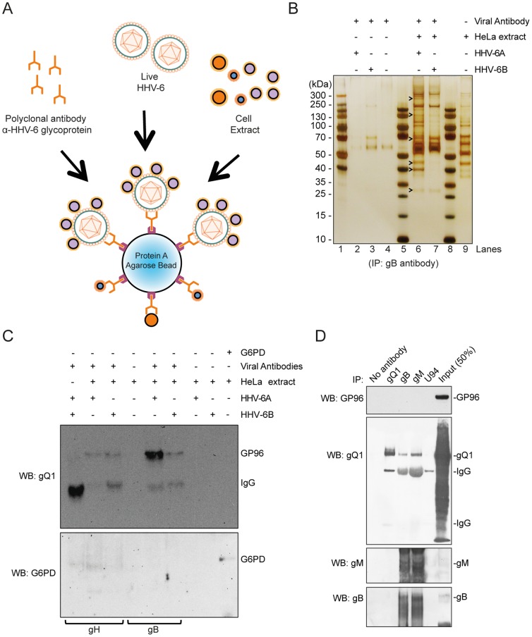
Figure 1. Identification of HHV-6-interacting host cell proteins.
(A) Diagrammatic representation of immunoprecipitation (IP) experiment to detect unknown HHV-6-interacting host cell proteins. (B) Protein complexes directly interacting with HHV-6 viral particles were isolated by IP using anti-HHV-6 gB antibody, separated by SDS-PAGE and visualized by silver staining. Molecular weight markers are indicated on the left. Arrowheads point to some of the proteins differentially identified by mass spectrometry. Lane 1, 5 and 8 shows pre-stained protein ladders. (C) IP and Western blotting were carried out to validate and characterize the interaction between GP96 and HHV-6 envelope glycoproteins. As negative control no antibody (Ab), no HHV-6, no HeLa lysate were used for IP. An antibody against human Glucose-6-P dehydrogenase (G6PD) in the absence of viral particles was also used as a control. The Western blot of the IP samples was probed for human GP96 and subsequently with G6PD. Anti-HHV-6 gB and anti-HHV-6 gH antibodies were used for IP. (D) Lysates of HSB-2 cells infected with HHV-6A were used for IP, followed by Western blotting (WB) to detect interaction between GP96 and HHV-6 glycoprotein complexes. The blot was probed with GP96, gQ1, gM and gB antibodies. IgG heavy and light chains are marked at appropriate places.

