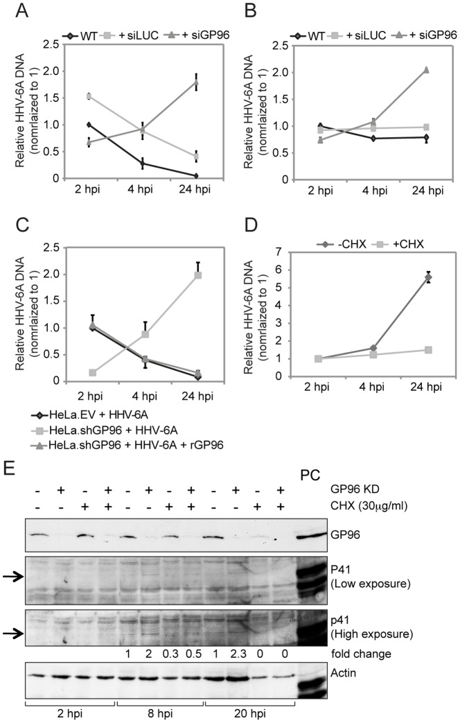
Figure 3. Absence of GP96 induces viral replication.
(A) Decrease in cell surface GP96 expression decreases HHV-6 entry but increases subsequent replication of viral DNA. HeLa cells were transfected with siRNA against human GP96. In parallel, cells were transfected with siRNA against the luciferase gene (siLUC) as a control. Blank transfected as well as siGP96 and siLUC transfected cells were washed thoroughly and infected with HHV-6A for 2 hrs. After 2 hrs of viral infection, cells were washed with trypsin and extensive washing with PBS to remove any bound but non-entered HHV-6 particles. Cells were then incubated for different time intervals in the absence of viral particle in the culture media and subsequently processed for quantification of viral DNA amount by qPCR. (B) The experiment described under (A) was performed in HSB-2 cells. (C) HHV-6A entry decreases in absence of GP96. HeLa cells carrying lentivirus-mediated stable knock down of GP96 (HeLa.shGP96) and vector control (HeLa.EV) were infected with HHV-6A. In parallel, HeLa.shGP96 cells were transiently transfected with a construct expressing human GP96 (rGP96) for 24 h and were also infected with HHV-6A. Cells were processed as mentioned above and amount of viral DNA in these cells was measured by qPCR. (D) Increase in viral DNA replication in the absence of GP96 requires host cell translation. HeLa.shGP96 cells were infected with HHV-6A in presence or absence of cycloheximide (CHX) and subsequently analyzed by qPCR. Data represent the mean ± SEM of three independent experiments. **, p≤0.005. Statistical analysis was based on the Student t-test. (E) Viral gene expression is increased in the absence of GP96. HeLa and HeLa.shGP96 cells were infected with HHV-6A for different time intervals in the presence or absence of CHX. Western blotting was carried out to detect HHV-6 early antigen p41. Actin served as loading control. 41 kDa bands specific for HHV-6 p41 is marked with arrow. HHV-6A infected HSB-2 cell lysate was used as a positive control (PC).

