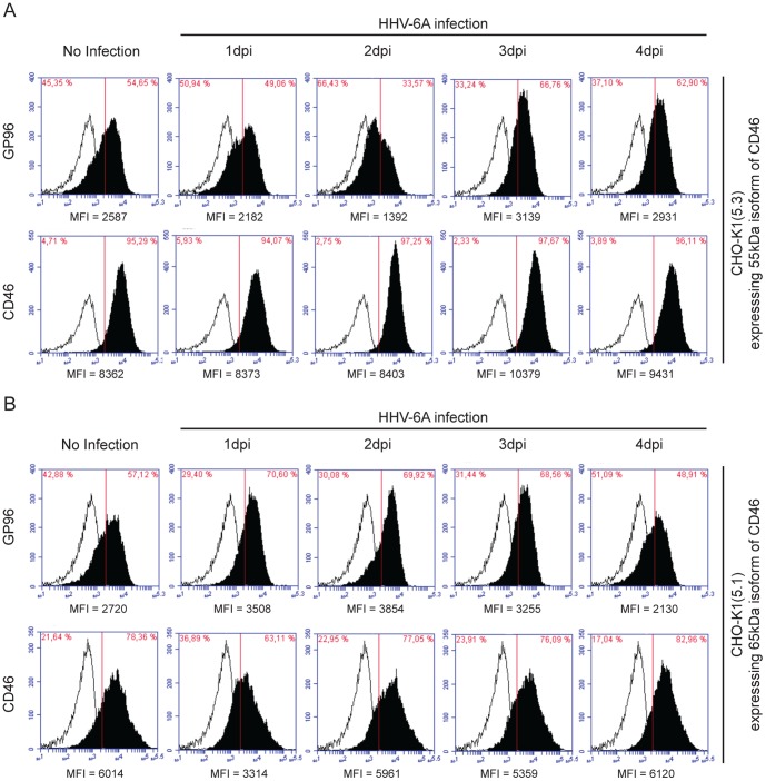
Figure 6. Association of CD46 with GP96 during HHV-6A infection.
(A) Cell surface expression dynamics of GP96 in CHO-K1 cells stably expressing 55 kDa isoform of CD46. CHO-K1(5.3) cells expressing the 55 kDa isoform of CD46 were infected with HHV-6A for indicated time points. CD46 and GP96 cell surface expression were analyzed by flow cytometry (solid profiles); background fluorescence levels were measured using an isotype specific antibody (empty profiles). (B) Cell surface expression dynamics of GP96 in CHO-K1 cells stably expressing 65 kDa isoform of CD46. CHO-K1(5.1) cells expressing the 65 kDa isoform of CD46 were infected with HHV-6A for indicated time points. CD46 and GP96 cell surface expression were analyzed by flow cytometry (solid profiles); background fluorescence levels were measured using an isotype specific antibody (empty profiles). Mean fluorescence intensity (MFI) for each analysis is indicated below the respective figure.

