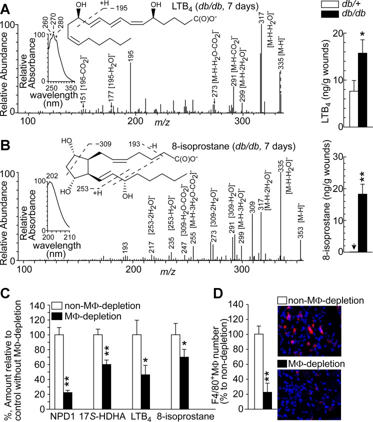
Fig. 2.
Higher levels of inflammatory LTB4 and oxidative-stress marker 8-isoprostane were observed after acute inflammation of wounds in diabetic db/db mice compared with non-diabetic db/+ mice. Macrophage (MΦ) depletion diminished the levels of NPD1, 17S-HDHA, LTB4, and 8-isoprostane in wounds. A: LTB4. B: 8-isoprostane. Left: aR chiral LC-MS/MS spectra with insets of aR chiral LC-UV spectra and MS/MS fragment interpretation. Right: LTB4 and 8-isoprostane levels in wounds collected at 7 dpw when wounds passed acute inflammation. C: amount (%), with MΦ-depletion, of NPD1 and 17S-HDHA in db/+ wounds or of LTB4 and 8-isoprostane in db/db wounds at 7 dpw relative to non-MΦ-depletion. D: MΦs in wounds at 7 dpw after depletion by Clodronate liposomes. Left: percentage of F4/80+ MΦs relative to non-depletion in wound sections. Right: immunohistological images. Data are means ± SE (n = 5). Significant difference: *P < 0.05; **P < 0.01.

