Fig. 3.
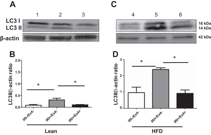
Ex4 mitigates the autophagy marker microtubule-associated protein 1A/1B-light chain 3 (LC3) II in liver of lean and HFD-fed mice exposed to IRI. After IRI and with or without Ex4 treatment, livers of lean (A and B) and HFD-fed (C and D) mice were homogenized, and equal amounts of protein were subjected to electrophoresis and probed with LC3 I/II antibody. A: Western blot for LC3 I/II and β-actin (loading control). Lane 1, control (IRI−); lane 2, IRI+/Ex4−; lane 3, IRI+/Ex4+. B: relative band density of LC3 II-to-β-actin ratio. Lean/IRI−/Ex4− vs. lean/IRI+/Ex4− (P < 0.04); lean/IRI+/Ex4− vs. lean/IRI+/Ex4+ (P < 0.04). C: Western blot for LC3 I/II and β-actin. Lane 4, HFD/IRI−/Ex4−; lane 5, HFD/IRI+/Ex4−; lane 6, HFD/IRI+/Ex4+. D: relative band density of LC3 II-to-β-actin ratio in HFD-fed mice. HFD/IRI−/Ex4− vs. HFD/IRI+/Ex4− (P < 0.01); HFD/IRI+/Ex4− vs. HFD/IRI+/Ex4+ (P < 0.002). Values are means ± SD; n = 9. *P < 0.05.

