
Fig. 4.
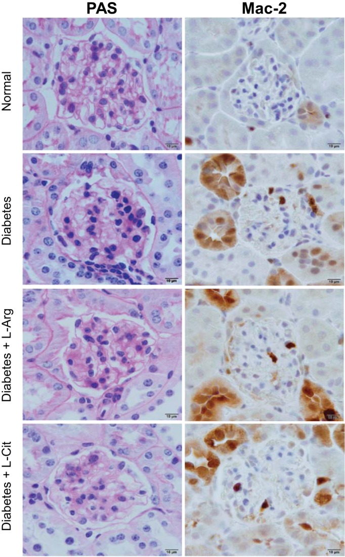
l-Arg or l-Cit supplementation did not improve renal histological changes or glomerular macrophage infiltration in diabetic mice. After 9 wk of diabetes with l-Arg or l-Cit supplementation as indicated, mice were euthanized and mouse kidneys were removed. Left: paraffin-embedded mouse kidney sections were performed with periodic acid-Schiff (PAS) staining, and images were taken with a ×100 (oil) objective with a total magnification of ×1,000. Semiquantitative scores (0∼4+) were assigned based on the masked readings, where 0 = no lesion and 1, 2, 3, and 4+ = mesangial matrix expansion or sclerosis involving ≤25%, 25∼50%, 50∼75%, or >75% of the glomerular tuft area, respectively. Right: glomerular macrophage recruitment was visualized by immunohistochemical staining using Mac-2 antibody at week 9 after STZ-induced diabetes.