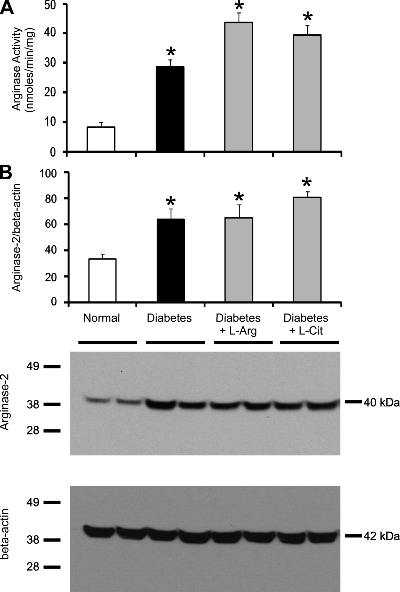
Fig. 6.
l-Arg or l-Cit supplementation did not affect kidney arginase activity or arginase-2 protein expression in diabetic mice. Mouse kidney tissue lysates were prepared after 9 wk of STZ-induced diabetes with or without l-Arg or l-Cit supplementation as indicated. Arginase activity (A) was measured as described in materials and methods. Western blot analysis was performed to detect arginase-2 protein. The membrane was then stripped and reprobed to measure β-actin as a loading control. Quantification was performed by densitometry followed by normalization to β-actin (B). Data are presented as means ± SE; n = 4–8 mice/group. *P < 0.05 compared with the normal group. A representative Western blot is shown in this figure and in subsequent figures.

