Fig. 5.
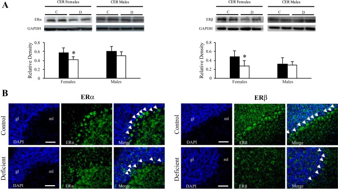
Localization of ERα and ERβ receptors in the CER of 21-day-old female rats. A: Western-blot analysis. Left: representative set of Western blots showing immunodetectable ERα protein. From left to right, the lanes contain total proteins isolated from CER (50 μg protein) of C and D 21-day-old rats. Right: representative set of Western blots showing immunodetectable ERβ protein. The lanes contain total proteins isolated from CER (50 μg of protein). Expression of both ERα (66 kDa) and ERβ (55 kDa) was quantified and normalized against GAPDH (38 kDa). Densitometric data were obtained from 5 separate experiments. Results are presented as means ± SD. Statistically significant differences between the 2 experimental groups: *P < 0.05 (ANOVA). B: immunohistochemical analysis. Observations were restricted in the cerebellar Purkinje cells of 21-day-old females exposed early to the deficient diet compared with control rats (n = 5/group). Localization of ERα (green; left) and ERβ (green; right) in the Purkinje cells of CER counterstained with DAPI (blue). Calibration bars, 200 μm. Original magnifications, ×20.

