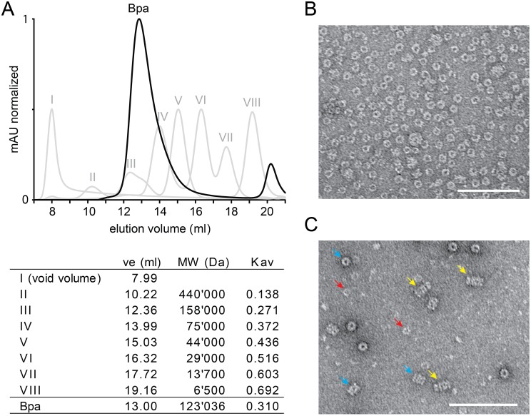
Figure 3. Bpa assembles into ring-shaped homo-oligomeric complexes and associates coaxially with the proteasome.
A, Analytical gel filtration profile of Bpa on a Superdex 200 column (black curve). Molecular size standards (I–VIII, grey curves) were run under the same conditions. The calculated molecular weight of Bpa at 13 ml elution volume is 123 kDa assuming an overall globular shape of the oligomer. B, Electron micrograph of negatively stained Bpa showing top views of the ring-shaped Bpa complexes at a magnification of 60′000. C, Electron micrograph of negatively stained Bpa and Δ7PrcAB shows top views of Bpa (examples indicated by red arrows) and Δ7PrcAB (turquoise arrows) as well as side views of the coaxially stacked complexes formed between Bpa and Δ7PrcAB (marked with yellow arrows). The scale bars in B and C both indicate 100 nm.

