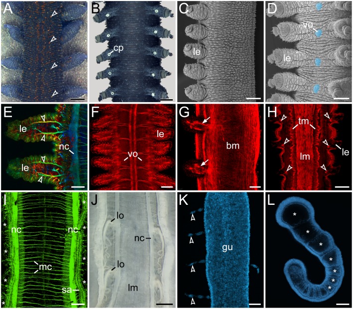
Figure 1. Segmental and non-segmental features in Onychophora.
(A, B, J) Light micrographs. (C, D) Scanning electron micrographs. (E–I, K, L) Confocal micrographs. (A) Trunk of a specimen of Euperipatoides rowelli in dorsal view. Note the segmentally repeated, terracotta-coloured papillae. (B) Trunk of a male E. rowelli in ventral view illustrating segmentally arranged crural papillae. (C) Trunk of a juvenile of Metaperipatus inae in lateral view. Note that segmentation is not evident in the integument. (D) Trunk of a juvenile of M. inae in ventral view. Segmentally arranged ventral/preventral organs between each leg pair are highlighted artificially in light-blue. (E) Legs of an embryo of Principapillatus hitoyensis in dorsal view. Anti-acetylated alpha-tubulin immunolabelling. Note the segmental arrangement of the anterior and posterior leg nerves (arrowheads). (F) Segmentally repeated anlagen of the ventral and preventral organs in an embryo of Epiperipatus biolleyi in ventral view. Phalloidin-rhodamine labelling of f-actin. (G) Segmental limb muscles (arrows) in an embryo of E. rowelli in lateral view. Phalloidin-rhodamine labelling. (H) Segmental arrangement of intrinsic leg muscles (arrowheads) in E. rowelli. Horizontal Vibratome section, phalloidin-rhodamine labelling. Note that the longitudinal and transverse musculature does not show any segmentation. (I) Ventral body wall of an embryo of P. hitoyensis in dorsal view. Anti-acetylated alpha-tubulin immunolabelling. Asterisks indicate the position of legs. Note that the median commissures are not arranged in a segmental fashion. (J) Enlarged modified nephridia ( = labyrinth organs) in the fourth and fifth leg-bearing segments. Note their segmental arrangement. Note also the lack of segmental ganglia associated with nerve cords. (K) Dissected midgut of a fully developed embryo of P. hitoyensis with segmentally arranged cellular strands (cf. [47]). DNA staining with Hoechst. (L) Embryo of P. hitoyensis in lateral view showing the segmental somites (coelomic cavities marked by asterisks). Subset of a confocal z-series showing an optical sagittal section. Abbreviations: bm, musculature of the body wall; cp, crural papilla; gu, midgut; le, leg; lm, longitudinal musculature; lo, labyrinth organ; mc, median commissures; nc, nerve cord; sa, salivary gland; tm, transverse musculature; vo, ventral/preventral organs. Scale bars: 1 mm (A), 500 µm (B), 300 µm (C–D), 50 µm (E), 100 µm (F, G, I, K, L), 200 µm (H), 1 mm (J).

