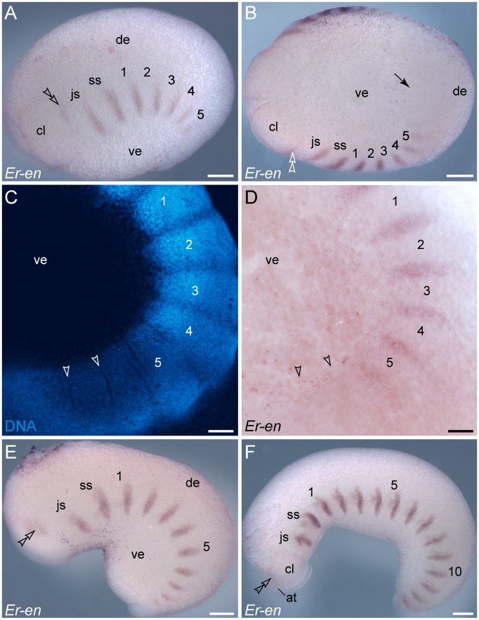
Figure 3. Expression of engrailed in embryos of the onychophoran E. rowelli at subsequent developmental stages.
Anterior is left in A, B, E, F and up in C, D. Leg-bearing segments and corresponding limbs are numbered. Note the repeated stripes along the body and the lack of expression in the dorsal and ventral extra-embryonic tissue. Double-arrowheads in A, B, E and F indicate the dorsally located domain of engrailed in the cephalic lobe. Arrowheads in C and D point to the transverse ectodermal furrows, which lack engrailed expression. (A) Early stage II embryo in lateral view. (B) The same embryo as in A in ventral view. Note the lack of expression in the posterior region and around the proctodaeum (black arrow). (C) Confocal micrograph of the posterior end of the same embryo as in A labelled with the DNA marker SYBR Green. (D) Light micrograph of the same embryo as in C. (E) Early stage III embryo in lateral view with eleven engrailed stripes along the body and a spot-shaped domain in the developing antenna (arrowhead). (F) Late stage III embryo in lateral view with 15 engrailed stripes along the body (13 within the trunk) and an additional domain in the developing antenna (arrowhead). Abbreviations: at, developing antenna; cl, cephalic lobe; de, dorsal extra-embryonic tissue; js, jaw segment; ss, slime papilla segment; ve, ventral extra-embryonic tissue. Scale bars: 250 µm (A, B, E, F), 100 µm (C, D).

